Blood clotting measures: understanding blood
A measure of blood clotting capacity may sound like dry laboratory jargon, but behind it lies a very practical question: can blood clot when it needs to while remaining fluid inside your blood vessels? Blood is more than just a red liquid: it is a transport, defense and regulatory system that keeps your tissues supplied with oxygen, nutrients and heat.
Here is a more readable overview of blood from part V of the physiology series: how much blood your body contains, what it consists of, how blood cells work, and the roles of blood plasma, blood groups, pH and gas transport. This text is for educational purposes and does not replace medical advice, especially on bleeding, anemia, clotting disorders or blood transfusions.
Measuring blood clotting capacity and the functions of blood
Blood accounts for approximately 6-9 % of human body mass. For a 70 kg person, this means around 5 l of blood—a normal blood volume, known as normovolemia. Losing 1/3 of your blood is life-threatening. An increase in blood volume is called hypervolemia, and a decrease is called hypovolemia.
Blood is a fluid connective tissue. Together with lymph and interstitial fluid, it forms the body’s internal environment. It has more than one function. Blood carries oxygen from the lungs to the tissues, delivers nutrients absorbed from the digestive tract to the cells, transports carbon dioxide back to the lungs and carries metabolic waste to the kidneys. Hormones and other biologically active substances also travel through the blood.
The simple truth is that without blood flow, neither oxygen nor glucose would reach your muscles. This becomes particularly clear during training, as working muscles need more oxygen and release more heat. Blood helps distribute this heat throughout your body and keeps temperature, osmotic pressure and pH as stable as possible.
White blood cells, antibodies and phagocytosis contribute to the blood’s protective function. Blood clotting is part of the same defense system: without it, you would bleed to death from even the smallest wound. This is where measuring blood clotting capacity comes in, because clotting must be sufficient without becoming excessive.
Blood: plasma, cells and measures of clotting capacity
Blood consists of plasma and formed elements, or blood cells. Plasma accounts for approximately 57% and blood cells for 43%. Plasma contains 92 % water, which gives blood its fluidity, 7-8 % protein and around 1 % low-molecular-weight substances, such as carbohydrates and lipids. Low-molecular-weight substances are present at approximately 20 g/L.
Inorganic substances, such as Na, K, Ca and Mg ions, affect the osmotic pressure of the plasma, and thereby the balance of water and salts. Organic substances in plasma include glucose, amino acids, fatty acids and lactic acid. Blood plasma proteins, present at around 65-80 g/l, participate in the exchange of water and substances between the blood and the tissues.
Albumin accounts for about 55 -60%, or approximately 60%, of plasma proteins. It helps maintain oncotic and osmotic pressure, influences blood volume and transports fatty acids, hormones, bilirubin, urobilin, bile salts, copper and certain substances foreign to the body, such as penicillin, in the blood. Albumin levels may fall with inflammatory diseases and damage to the liver or kidneys.
Globulins are a broad group of proteins involved in the transport of lipids, cholesterol, copper and iron. Around 2/3 of plasma glucose may be bound to glycoproteins. Globulins also include immunoglobulins, or antibodies, and the red blood cell-agglutinating substances anti A and anti B. The original material also mentions a B11-binding globulin.
Fibrinogen is a plasma protein central to blood clotting. At the end of a cascade of reactions, dissolved fibrinogen becomes fibrin, which forms the fibrous framework of a clot. Plasma fibrinogen levels are therefore directly linked to what a measure of blood clotting capacity means in practice.
Anemia, polycythemia and changes in white blood cells
Anemia primarily means a reduced oxygen-carrying capacity of the blood due to a lack of hemoglobin. Iron deficiency anemia is a common form. It may be caused by insufficient iron in the diet, impaired iron absorption from the digestive tract or chronic blood loss, for example from gastrointestinal ulcers, carcinomas, polyps, diverticula or heavy menstrual bleeding.
A key feature of megaloblastic anemias is the presence of abnormally large red blood cells and their immature precursors in the blood and bone marrow. Hypochromic microcytic anemia, hemolytic anemia, aplastic anemia and pancytopenia are also mentioned. Polycythemia means an increase in red blood cell count and hemoglobin concentration.
Leukocytosis is an elevated white blood cell count in circulating blood. It may occur with infectious diseases, inflammatory processes, tissue necrosis, brain injury or after the use of certain medications. Leukopenia means a low white blood cell count. Leukemia is a cancerous process arising in a blood-forming stem cell—a malignant disease that affects the blood-forming organs.
Blood cells and oxygen transport
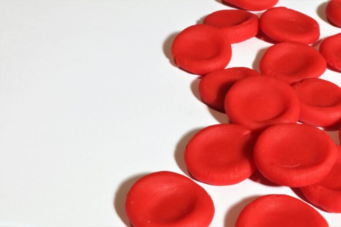
Blood cells fall into three types: red blood cells, white blood cells and platelets. Erythrocytes number approximately 4.5–5 million/mm³, leukocytes 4,000–10,000/mm³ and platelets 150,000–300,000/mm³. Their counts provide a great deal of information about the body’s condition. An older method involves diluting the blood in a fixed ratio and counting the cells under a microscope; electronic cell counters are also used today.
Erythrocytes are cells without a nucleus, with a diameter of approximately 7.5 Mm and a lifespan of 100-120 days. Women have fewer on average than men: men – 40 -51 % and women – 36 – 47 %. The erythrocyte sedimentation rate can help assess inflammatory diseases, as inflammation causes the cells to settle faster.
Hemoglobin makes up 15% of blood. Hemoglobin consists of 4 heme groups and 4 globin chains and contains 4 Fe 2+ ions that bind oxygen. In simplified form, the reaction can be written as follows: Hb +4O2 Hb (O2)4. The average hemoglobin concentration is 158 g/l in men and 140g/l in women. 1 mole of hemoglobin can bind a maximum of 4 moles of oxygen.
In practical terms, 1 g of hemoglobin binds 1.39 ml O2; values of 1.34 to 1.36 ml/g are also used in blood gas analysis, while 1.34 ml O2 is often used for in vivo calculations. Oxygen binding is affected by the partial pressure of O2, temperature and pH. In working muscle, temperature rises and pH may fall, promoting the release of oxygen from hemoglobin.
Oxyhemoglobin is an unstable compound formed when oxygen binds to hemoglobin. The oxyhemoglobin dissociation curve describes the relationship between HbO2 and pO2. Point A represents the relationship between pO2 and HbO2 % in arterial blood, while point B represents the same relationship in venous blood. The curve rises steeply at low pO2 values and more gradually at high pO2 values.
If the O2 content of arterial blood is 19% by volume, it may fall to 11% in venous blood draining from the tissues. This means that 8% by volume of oxygen has been delivered to the tissues. This difference is called the arteriovenous oxygen difference. It indicates how much oxygen each 100 ml of blood delivers to the tissues.
Leukocytes and defense
Leukocytes are cells with a nucleus and a diameter of 7.5 – 20 Mm. Their lifespan can range from 12 h to several years. Granulocytes live for 12 h to 2 days, number approximately 4900 per 1 mm3 and account for 50-70% of leukocytes. Neutrophilic granulocytes are important phagocytes, eosinophilic granulocytes strengthen the body’s defenses, and basophilic granulocytes can produce large amounts of histamine.
Monocytes account for 4-8 % of leukocytes and have a strong capacity for phagocytosis. Neutrophils tend to attack smaller cells, while monocytes attack larger ones. They move by amoeboid movement and can leave blood vessels to reach sites of inflammation.
Agranulocytes can live for weeks to years. They number approximately 4000 per 1 mm3 and account for 30-50% of leukocytes. Lymphocytes are an important part of the immune system. In adults, they account for approximately 25 to 40 % of all leukocytes. They are divided into T and B cells, with T cells accounting for approximately 80 %.
When a foreign protein enters the body, lymphocytes begin to multiply and become more active. On first exposure, activation may take a week or more. On subsequent exposure, the response is faster because the immune system already recognizes the foreign substance.
Blood clotting capacity indicator and platelets
Platelets are small structures without a nucleus. They have a diameter of 1-4 Mm and a lifespan of 5-11 days; the overview of blood cell formation also mentions 10 days. Platelets contain factors essential for blood clotting and substances that constrict blood vessels, as well as adrenaline and other compounds.
Blood clotting begins when a blood vessel is damaged. Platelets release vasoconstrictors, which narrow blood vessels at the injury site. Platelets stick to one another and to the damaged area. A white thrombus forms, reducing bleeding. At the same time, a red thrombus begins to form.
The main reaction in red thrombus formation is the conversion of the plasma protein fibrinogen into insoluble fibrin by thrombin. Approximately 20 clotting factors are involved in blood clot formation. To understand a blood clotting capacity indicator, you need to consider the whole process: platelets, clotting factors, fibrinogen and the blood vessel wall work together.
- Activation phase: platelets adhere to the damaged area of the blood vessel, and prothrombin activation begins.
- Coagulation phase: prothrombin is converted into thrombin, which releases fibrin from fibrinogen and creates the clot’s framework.
- Retraction phase: the clot is replaced by connective tissue, with platelets also contributing to this process.
- Fibrinolysis phase: the clot may later dissolve, allowing blood to flow through the vessel again.
Anticoagulants inhibit clotting. The original material lists a drop in temperature, non-wettable surfaces, Na oxalate, K oxalate, ammonium oxalate, Na citrate, EDTA, heparin and coumarin derivatives among these. Other anticoagulants mentioned include hirudin, antithrombin, certain snake venoms and tabanin.
Blood cell formation and lifespan
Blood cell formation takes place mainly in the red bone marrow. The main organs involved are the red bone marrow, lymph nodes, thymus and spleen. Red bone marrow is found in the sternum, ribs, skull, pelvis and proximal portions of the long bones. All blood cells are produced there.
The lymph nodes, thymus and spleen are primarily involved in the maturation of lymphocytes and monocytes. Blood plasma proteins are produced in the liver, although the liver is not strictly considered a blood-forming organ. Red bone marrow contains pluripotent stem cells that can develop into different types of blood cells.
Red blood cells have a lifespan of 100-120 days. Among white blood cells, granulocytes may be short-lived, lymphocytes can live for years, monocytes for weeks, basophils for 12 hours and neutrophils for 6-8 hours. Platelets have a lifespan of about 10 days.
Erythropoiesis, the production of red blood cells, is influenced by vitamin B 12, folic acid and iron. Iron is needed to build hemoglobin; without hemoglobin, a red blood cell cannot fulfill its role of carrying oxygen. The kidneys play a key role in the synthesis of erythropoietin, and its production depends on the body’s oxygen supply. A lack of O2 triggers erythropoiesis.
After approximately ~120 days in circulation, red blood cells are broken down by cells of the reticuloendothelial system, primarily in the bone marrow and, in pathological conditions, also in the liver and spleen. As hemoglobin breaks down, its porphyrin component is converted into bilirubin and excreted into the intestine in bile. Iron (2+) returns to the plasma bound to transferrin, where it is present as iron (3+). Approximately 18% of the iron released is reincorporated into the heme structure.
Blood groups, the Rh factor and blood transfusion
In the ABO system, blood groups are distinguished by the antigens on the surface of red blood cells and the antibodies in blood plasma. The A antigen must not come into contact with anti-A antibodies, nor the B antigen with anti-B antibodies. Otherwise, red blood cells clump together, hemolysis occurs and the transport of respiratory gases is impaired.
- 0 (I): genotype 00, anti-A and anti-B antibodies in the plasma.
- A (II): genotype A0 or AA, A antigens on red blood cells, anti-B antibodies in the plasma.
- B (III): genotype B0 or BB, B antigens on red blood cells, anti-A antibodies in the plasma.
- AB (IV): genotype AB, A and B antigens on red blood cells, no antibodies.
The D antigen is the most important antigen in the Rh system. If red blood cells carry the D antigen, the blood is Rh-positive. If the D antigen is absent, the blood is Rh-negative. About 85 % of people have Rh-positive blood. A person with Rh-negative blood must not receive Rh-positive blood if anti-D antibodies could trigger agglutination and hemolysis.
Rh incompatibility can also occur during pregnancy if the mother is Rh- and the father is Rh+, and the baby is Rh+. The first pregnancy is usually not a problem because anti-D antibodies are not present in the body at birth; they develop following exposure to Rh+ blood, for example during childbirth or an abortion. During the second pregnancy, anti-D antibodies can cross the placental barrier and cause agglutination in the fetal circulation.
In Central Europe, more than 40 % of people have blood group A, 40 % have group O, well over 10% have group B and ~6 % have group AB. In cases of minor blood loss, a person with blood group 0- is considered a universal donor. Larger transfusions must use blood of the same group. People with blood group AB lack the corresponding antibodies in their plasma, so they can receive small quantities of blood from other groups.
Blood pH, buffers and oxyhemoglobin
Blood pH indicates the amount of hydrogen ions present. The more free H ions there are, the more acidic the environment and the lower the pH value. Arterial blood plasma has a pH of about 7.4, while venous plasma has a pH of 7.35. Blood pH varies within the limits of 7.37 to 7.43. The pH inside red blood cells is lower, at about 7.2 to 7.3.
Acidosis means a fall in blood pH, or an increase in acid equivalents, pH < 7.37. Alkalosis means a rise in pH, pH >7.43. Both can disrupt enzyme activity and metabolism. Relative to water, the average viscosity of blood in healthy adults is 4.5, and that of blood plasma is 2.2. An increase in hematocrit increases viscosity. A healthy man has a hematocrit of 0.44-0.46, and a healthy woman 0.41-0.43. In newborns it is 20% higher, and in young children 10% lower, than in women.
The body also produces acidic compounds during normal metabolism. Glycolysis produces lactic acid and free H ions. Aerobic oxidation of glucose produces CO2. Incomplete oxidation of fatty acids produces ketone bodies. The breakdown of phosphates and sulfur-containing amino acids also contributes.
- Bicarbonate buffer: based on CO2 and bicarbonate and depends heavily on respiration.
- Phosphate buffer: consists of monobasic phosphate H2PO4 and dibasic phosphate HPO4, but has a smaller effect in the blood.
- Protein buffer: relies on plasma proteins, particularly albumin, and amino acid side chains.
- Hemoglobin buffer: very important because hemoglobin is abundant and its acidity changes depending on whether it is bound to oxygen.
Oxyhemoglobin is more acidic than deoxygenated hemoglobin within the physiological pH range. O2 exchange therefore enhances hemoglobin’s buffering capacity. Breathing helps regulate pH by removing CO2: when acidity increases, hyperventilation can expel more CO2, while increased alkalinity reduces ventilation. The kidneys help excrete nonvolatile acids, such as sulfuric acid.
Transport of substances in the blood
Oxygen travels through the blood mainly bound to hemoglobin, but before entering a chemical combination, every O2 or CO2 molecule must briefly be physically dissolved. In the lungs, the partial pressure of oxygen is higher, and hemoglobin binds oxygen. In the tissues, the partial pressure of O2 is lower, and hemoglobin releases oxygen.
CO2 transport is more complex. Blood carries carbon dioxide dissolved in blood plasma and electrolytes, given in the original as VP 5% and E 7%; bound to proteins, primarily hemoglobin in red blood cells and to a small extent proteins in plasma, 11%; as bicarbonate in blood plasma and red blood cells, VP 94% and E 82%; and in very small amounts as undissociated carbonic acid.
Gas exchange in the tissues occurs by diffusion through capillary walls, driven by differences in partial pressure. As O2 dissolved in blood plasma moves into tissue fluid and then into cells, blood pO2 falls. Hemoglobin responds by releasing more oxygen, shifting the balance toward meeting the tissues’ needs.
Practical signs of blood-related problems to watch for
If you exercise, you may notice blood-related issues indirectly. Fatigue, shortness of breath, paleness, lower exercise tolerance than usual or prolonged bleeding do not establish a diagnosis on their own, but they are reasons to speak to your family doctor. The same applies if your blood test shows hemoglobin, white blood cells, platelets or clotting-related markers outside the normal range.
Training does not automatically make you healthier if your recovery, sleep and nutrition are poor. Deficiencies in iron, vitamin B 12 and folic acid can affect red blood cell production. Dehydration, in turn, can affect blood volume and how you feel. Frankly, it’s not glamorous, but it’s the essential groundwork.
If your doctor says a blood clotting indicator is problematic, you should not try to correct it yourself with supplements or random advice. The clotting system depends on balance: too little clotting increases the risk of bleeding, while too much increases the risk of thrombosis. Both require careful assessment.
FAQ: blood clotting capacity indicators
What is a blood clotting capacity indicator?
This is a general way of describing how well blood can clot after an injury. Physiologically, this depends on platelets, clotting factors, fibrinogen, the blood vessel wall and the balance of anticoagulants.
Why is oxyhemoglobin important?
Oxyhemoglobin is hemoglobin bound to oxygen. It enables oxygen to be transported from the lungs to the tissues and also contributes to the blood’s buffering capacity, because hemoglobin’s properties change when it binds oxygen.
Does a change in blood pH affect training?
Yes, because enzyme function and muscle metabolism depend on pH. The body keeps blood pH within a narrow range of 7.37–7.43 through buffer systems, breathing and kidney function.
Author: EKFK
Come and work out! ArtGym
Visit the marketplace! https://turg.fitness.ee/et