Digestion
General overview of the digestive tract and digestive processes.
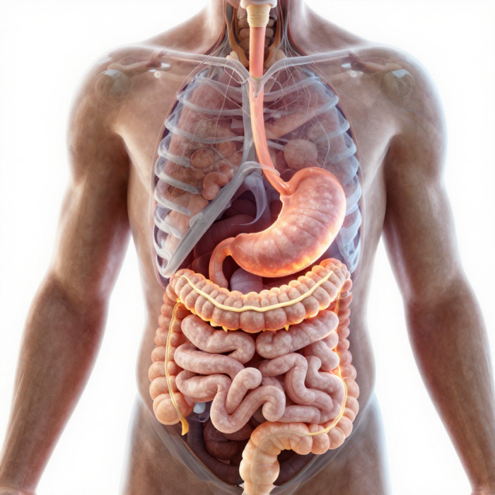
Main parts of the digestive tract: Oral cavity, esophagus, stomach, duodenum, small intestine, large intestine.
Glands producing digestive juices and their secretions. Mouth: 3 pairs (parotid, sublingual and submandibular glands) plus many small salivary glands in the oral lining – saliva. Stomach: gastric mucosal glands – gastric juice. Small intestine: liver glands – bile; pancreas – pancreatic juice; small intestinal glands – intestinal juice. Large intestine –
Digestive processes: mechanical, secretory and absorption-related phenomena. Food is processed mechanically in the digestive tract and mixed with digestive juice, whose enzymes break nutrients down so they become absorbable and acceptable to the body; proteins and lipids lose their species specificity.
Approximate time food takes to pass through the digestive tract. Food stays in the oral cavity for 15-18 seconds. Solid food travels from the oral cavity to the stomach in 8-9 seconds, liquid food in 1-2 seconds. Food stays in the stomach for 4-10 hours. It does not stop in the duodenum. Final digestion takes place in the small intestine, 10-15 hours.
In the large intestine, 1-2 days go to moving waste onward.
Digestion in the oral cavity
Food stays in the oral cavity for no more than 15-18 seconds. Even so, its mechanical (moistening with saliva, grinding) and chemical (salivary enzymes acting on complex sugars) processing begins here, making it swallowable. The oral cavity is also where food is assessed, i.e. its taste and edibility are determined. In the oral cavity food is ground with the teeth and mixed with the tongue.
Saliva is a slightly alkaline fluid (pH 7.4-8.0) containing 98-99% water and 1-1.5% salts and organic matter. Its enzymes include amylase, which breaks down carbohydrates, and lingual lipase, which breaks down lipids and comes from Ebner’s glands at the base of the tongue.
The average daily amount of saliva is 1-1.5 l, depending on the composition and water content of the food.
Besides amylase and maltase, saliva contains other organic and inorganic substances. An example of the organic ones is the protein compound mucin, a mucous substance that makes the bolus of food slipperier and eases swallowing. Lysozyme kills bacteria.
Relative amount and composition of saliva produced by the different salivary glands.
The parotid glands secrete thin (serous) saliva. The secretion of the sublingual and submandibular glands is rich in mucus.
Reflex regulation of salivary gland function. The main stimuli are taste and purely mechanical touch. The centers in the CNS (in the medulla oblongata and the hypothalamus, which is in turn controlled by the cerebral cortex) play a major role. When something is put into the mouth (taste), receptors send signals to the salivation center (in the medulla oblongata).
Olfactory receptors send signals to the salivation center of the hypothalamus.
Conditioned reflex link – e.g. the mouth waters at the clatter of dishes.
The digestive tract is controlled by the autonomic nervous system.
The sympathetic and parasympathetic nerves are equally stimulating: parasympathetic stimulation produces plenty of thin saliva, sympathetic stimulation a thicker, more enzyme-rich saliva.
The oral cavity as a reflexogenic zone. A mouthful of something good is enough, and the whole digestive tract is ready to receive food. Swallowing is reflexive. Chewed food, moistened with saliva, moves to the base of the tongue, and by moving the tongue back and up it is pushed through the pharyngeal isthmus into the pharynx. As food passes through the pharynx, its path crosses the airways: the soft palate closes off the nasal cavity from behind, the larynx is raised under the base of the tongue and covered by the epiglottis, breathing stops reflexively, and the food enters the esophagus, where wave-like peristaltic contractions of its muscles carry it on to the stomach. Solid food covers this distance in 8-9 seconds, liquid food in 1-2 seconds.
Digestion in the stomach
Gastric motility ensures that food is received, mixed with gastric juice, and that the stomach empties. Food is digested in the stomach for 4-10 hours. Further mechanical and chemical processing take place here. Mechanical processing (churning, rubbing, kneading) is provided by gastric motility – contractions of the strong smooth muscles in the stomach wall. The result is pulsation (mixing, so that the enzymes can reach the food). Chemical processing takes place under the influence of gastric juice, secreted by glands in the gastric mucosa. Gastrin formed in the G cells of the gastric mucosa, and motilin and cholecystokinin from the small intestinal mucosa, stimulate gastric motility, while secretin, gastric inhibitory polypeptide (GIP) and glucagon inhibit it.
A person produces 1.5-2.5 l of gastric juice per day. Gastric juice contains enzymes, hydrochloric acid and mucus.
The main enzymes in gastric juice are proteases and lipase. Proteases include the pepsins, which the glandular cells of the stomach release as inactive pepsinogens.
Pepsinogen is activated by HCl (hydrochloric acid). Pepsins break down dietary proteins into polypeptides.
Gastric juice contains only a little lipase, because the stomach environment is unsuitable for it (gastric juice pH 1.5-3.5, its optimum 5.0). Lipases are effective with emulsified fats (e.g. milk fat).
Lysozyme (killing bacteria) and mucin (lubricating food) matter as much in the stomach as in the oral cavity. In the stomach, mucin also has a very important function for the stomach wall (protecting it against physical and chemical influences); it is a glycoprotein and tends to bind vitamins (protecting them from stomach acids, which would otherwise destroy them and they would simply be lost).
The acidic reaction of gastric juice comes from the hydrochloric acid it contains, which is important because it:
1. activates pepsinogens; 2. denatures and swells proteins, which promotes their enzymatic breakdown; 3. promotes the curdling of milk, which is necessary for its digestion
4. activates the hormone gastrin, which is produced by the mucosa of the pylorus and increases gastric juice secretion; 5. on entering the duodenum, takes part in the formation of many hormones that regulate stomach, pancreas and liver function; 6. stops putrefactive processes in the stomach; 7. promotes gastric motility; 8. takes part in the complex mechanism by which food masses pass from the stomach into the intestine.
In the regulation of gastric juice secretion 3 phases can be distinguished: 1. the brain, or cephalic phase;
2. the stomach, or gastric phase; 3. the intestinal phase. These phases partly overlap in time.
CEPHALIC PHASE. The sight, smell and taste of food, followed by chewing and swallowing, trigger gastric juice secretion. This rests on unconditioned reflexes, whose afferent pathway can vary; the efferent part runs via the vagus nerve and reaches the parietal, i.e. G cells through the enteric NS. The main transmitter at the nerve endings is acetylcholine. Gastric juice secretion intensifies under the influence of conditioned or unconditioned reflexes, i.e. it is a very fast regulation mechanism (parasympathetic excitation). THE GASTRIC PHASE begins as soon as food enters the stomach (within 30 minutes of starting to eat); secretion is stimulated by both physical (stretch) and chemical (peptides, amino acids, alcohol, caffeine etc.) stimuli. When food reaches the stomach, the acidity of the stomach contents decreases, which is one of the factors triggering gastrin. THE INTESTINAL PHASE begins when food reaches the duodenum. The stimuli are stretching of the small intestinal wall, the chemical composition of the food and osmotic pressure… Stimulating the mechanosensors inhibits gastric motility and raises the tone of the gastric sphincter; higher osmotic pressure triggers GIP release via osmosensors, which inhibits gastric juice secretion and motor activity. The chemosensors of the duodenum monitor the pH of the chyme; when the intestinal contents become more acidic, secretin is released. Secretin stimulates the secretion of bicarbonate-rich pancreatic juice, while inhibiting gastric juice formation and lowering gastric motility. Amino acids reaching the duodenum trigger the release of cholecystokinin (CCK), which causes the production of enzyme-rich pancreatic juice and gallbladder contraction and intensifies intestinal motility.
The secretory function of the stomach takes place in 2 phases: 1. IN THE REFLEX PHASE gastric juice is secreted under the influence of conditioned stimuli (the look and smell of food, indifferent stimuli that previously coincided with receiving food) and unconditioned stimuli (mechanical irritation of the mucous membrane of the mouth and pharynx by food). The excitation arising in the receptors is passed on to the nutrition center, from where impulses travel along the efferent fibers of the vagus nerve to the gastric glands. Conditioned reflex secretion of gastric juice can begin at the smell and sight of food. This juice is called appetite juice, and its secretion prepares the stomach to receive food. Appetite, i.e. the sensation of needing to take food into the body, promotes excitation of the gastric glands. Appetite arises through the influence of conditioned and unconditioned stimuli. That is why eating conditions and the taste of food matter in digestion. Still, despite the large role of conditioned reflex stimuli in the onset of appetite, a change in blood composition is considered the main stimulus of the nutrition center. 2. THE NEUROHUMORAL PHASE begins 30 minutes after receiving food. In this phase, many factors stimulate gastric secretion. Mechanical irritation of the stomach wall by food masses triggers gastric gland secretion by reflex and chemical routes. Under mechanical irritation, the mucosa of the pylorus produces the hormone gastrin, which, absorbed into the blood, promotes gastric gland function. Chemical irritation of the duodenum likewise leads to the formation of a hormone in its mucosa, which triggers juice secretion in the stomach. In addition, histamine (formed in the body’s tissues and also contained in food), protein digestion products, and the extractives in meat and root vegetables take part in the chemical stimulation of the gastric glands.
The effect of different nutrients and the hormone gastrin on gastric function. Absorbed into the blood, gastrin promotes gastric gland function. Histamine (formed in the body’s tissues and also contained in food), protein digestion products, and the extractives in meat and root vegetables take part in the chemical stimulation of the gastric glands.
Regulation of gastric motor function – the role in it of the autonomic nervous system, the chemo- and mechanoreceptors of the gastric mucosa, gastrin and catecholamines.
Digestion in the small intestine
Pancreatic juice is alkaline, pH 7.8-8.4. It contains enzymes that break down proteins (trypsin etc.), polypeptides, fats (lipase) and carbohydrates (amylase, maltase etc.). Pancreatic juice contains HCO and enzymes for breaking down all nutrients. About 1.5 liters are produced per day.
The main enzymes in pancreatic juice and their role in digestion. LIPASES – breaking down dietary fats into glycerol and fatty acids (a pH-favorable environment), working effectively in the presence of bile. AMYLASE, MALTASE, LACTASE, SUCRASE – action on carbohydrates. NUCLEASES (ribonucleases) – breaking down nucleic acids. PROTEOLYTIC ENZYMES (endopeptidases) – analogous to pepsin (secreted inactive and made active in the 12-finger intestine (duodenum), so that they do not start damaging the zones that produce them). Endopeptidases – breaking down the protein molecule from within (trypsin, chymotrypsin). CARBOXYPEPTIDASES start the breakdown from the carboxyl group. AMINOPEPTIDASES – start the breakdown from the amino group. DIPEPTIDASES – these must be present.
In the activation of proteolytic enzymes the enzyme enterokinase plays an important role; it is found in the juice produced by the duodenum. Proteolytic enzymes are secreted inactive so that they do not start damaging the zones that produce them.
Bile is produced continuously in the liver, and the daily amount can reach 0.5-1 liter (about 400 ml). Bile contains no enzymes. It is a golden-yellow fluid (golden-brown to greenish) that contains bile acids, bilirubin and other substances.
Functions of bile in digestion. Bile strongly activates the lipases in pancreatic juice and intestinal gland juice. It emulsifies dietary fats (lipids) (so that they can be absorbed so easily), affects the condition of the intestinal wall (making it highly permeable to fats and fatty acids), and as a result promotes the absorption of fat-soluble vitamins, clearly stimulates intestinal motility, etc.
Amount and composition of intestinal juice, main enzymes found in intestinal juice.
The small intestinal glands produce about 1.5 l of intestinal juice per day; it is close in character to extracellular fluid (pH 6.5-7.5) and contains enzymes that break down disaccharides: maltase, sucrase and lactase. Intestinal juice is produced in the Lieberkühn glands and contains plenty of a mucous substance that protects the intestinal wall.
On the brush border of the digestive tract there are peptidases, lipase, maltase, lactase, sucrase and other enzymes from pancreatic and intestinal juice, because intestinal epithelial cells actively absorb digestive enzymes. This is where absorption takes place.
Pancreatic juice secretion is governed by neural and humoral factors. It arises through conditioned and unconditioned stimuli. Conditioned reflex secretion begins at the sight and smell of food, and in humans even when food is merely talked about. During eating, the receptors of the oral cavity and pharynx are mechanically stimulated. From there signals travel to the medulla oblongata and trigger pancreatic juice secretion via unconditioned reflexes. The secretory nerves of the pancreas contain fibers of the vagus nerve. The chemical trigger of pancreatic secretion is the hormone secretin. It forms in the cells of the duodenal mucosa as inactive prosecretin, which is activated by the hydrochloric acid of gastric juice. Secretin is absorbed into the blood and stimulates pancreatic juice secretion. In the liver bile is produced without interruption, and the process intensifies during digestion. From the liver bile goes to the gallbladder, where it stays until food is taken in. The increase in bile production during digestion and the release of bile from the gallbladder into the intestine are governed by neural and humoral factors. The look and smell of food, the act of eating, and stimulation of the stomach and duodenal receptors by food masses intensify bile production and cause bile to be released into the intestine via conditioned and unconditioned reflexes. The secretory nerve of the liver is the vagus nerve. The sympathetic nerve reduces bile production and halts evacuation from the gallbladder. The chemical stimuli of the liver are digestive hormones (secretin), protein digestion products and extractives. Salts of bile acids promote strong bile secretion; absorbed into the blood, they stimulate the function of liver cells.
The wall of the small intestine is covered with epithelium, between which the tubular ducts of the Lieberkühn glands open. These glands secrete intestinal juice, which consists of a liquid part and denser mucus flakes suspended in it, produced mainly in response to unabsorbable substances that have entered the intestine and could damage the mucosa. The main factors stimulating the secretion of the intestinal glands are mechanical and chemical stimuli of the intestinal wall. Among the chemical stimuli, gastric juice, protein breakdown products, seasonings and milk sugar are of greater importance. The mechanical and chemical stimuli that stimulate intestinal juice secretion act on the local nervous apparatus of the intestine.
Absorption of nutrients in the small intestine. In the intestine, nutrients are absorbed into the blood and lymph. The nutrients the body needs become available to tissues and organs only after they have entered the blood from the digestive tract. Nutrient absorption takes place mainly in the small intestine. In other parts of the digestive tract only a few substances are absorbed, in small amounts (stomach – mineral salts, monosaccharides, alcohol, water; large intestine – water). Intensive absorption in the small intestine is ensured by its large surface area, which is increased by the villi, outgrowths of the mucosa. Inside each villus are smooth muscle fibers and a well-developed network of blood and lymph vessels. The villi are covered with epithelium. Intensive (parietal) digestion takes place on the surface of the microvilli. Absorption is a complex physiological process involving diffusion, filtration and osmosis. The intestinal epithelium is not merely a semipermeable membrane but also performs a secretory function, i.e. it ensures the selective absorption of one substance and restricts that of others. Proteins are broken down in the digestive tract into amino acids and pass into the blood in this form. Carbohydrates are absorbed mainly as glucose, which is explained by the ability of the intestinal wall to let this monosaccharide through selectively. Fats are absorbed from the small intestine as fatty acids and glycerol. The latter dissolves well in water and is easily absorbed through the intestinal mucosa. Fatty acids are absorbed only in the presence of bile, because they do not dissolve in water. Water can be absorbed already in the stomach, but most of it passes into the blood from the small and large intestine.
Digestion in the large intestine
Intestinal motility.
The role of large intestine juice and the microflora of the large intestine in digestion. Digestion of food mainly ends in the small intestine. The glands of the large intestine mucosa secrete a digestive juice that is relatively poor in enzymes. It contains small amounts of low-activity enzymes that break down residues of proteins, fats and carbohydrates. Juice secretion in the large intestine takes place under the influence of local stimuli of its mucosa.
The large intestine actively reabsorbs Na, with water following by diffusion driven by osmotic pressure. Extensive water reabsorption takes place in the large intestine. Under the action of bacteria, the unabsorbed amino acids and other protein digestion products are broken down in the large intestine. In the process, substances toxic to the body are formed; these are absorbed into the blood and the liver renders them harmless.
Smooth muscle tissue and striated muscle tissue in the digestive tract. The muscle tissue in the upper parts of the digestive tract is skeletal muscle tissue, from the middle of the esophagus onward it is smooth muscle tissue, and in the anal region there is skeletal muscle tissue under voluntary control.
The intestinal wall contains longitudinal and circular muscle layers. The circular muscle layer is the inner one and the longitudinal muscle
layer the outer one.
Four types of movement can be distinguished in intestinal motility. PENDULAR MOVEMENTS result from alternating rhythmic contractions of the circular and longitudinal muscle layers of the intestine. In a certain short segment the intestine alternately contracts and relaxes, and the intestinal contents move now in one direction, now in the other. Because different parts of the intestine do not contract at the same time, the contents undergo rhythmic segmentation – at one moment they are divided into separate portions, and then they merge again. Pendular movements ensure that the intestinal contents are mixed with digestive juices. PERISTALTIC MOVEMENTS consist in the fact that contraction of the circular muscles and the resulting narrowing of the intestine in one section is accompanied by relaxation of the muscles and widening of the intestine in the neighboring section located more distally. As a result, the contents shift from the narrowed section into the lower, widened section. At the next moment the muscles contract in the relaxed region of the intestine, while in the neighboring, more distal section they relax. Peristaltic movements ensure that the contents advance along the intestine toward the rectum. In the small intestine, segmentation and pendular movements.
In small intestinal motility villus movements can be distinguished, which promote contact of the intestinal contents with the intestinal epithelium and the absorption of nutrients. The chyme is mixed by the (non-propulsive) segmentation and pendular movements of the muscle layers of the intestinal wall. The contents are transported onward by (propulsive) peristaltic waves. Peristaltic waves manifest in the following way: when the longitudinal muscle layer of the intestinal wall contracts, the lumen of the intestine widens, and the contraction of the circular muscle layer immediately following it pushes the contents onward. For the large intestine, read the previous answer!
Regulation of intestinal motility: the influence of the autonomic NS, acetylcholine, noradrenaline, and the chemical and mechanical properties of the food mass. The nerves supplying the digestive tract come from the sympathetic and parasympathetic parts of the autonomic NS, and influences transmitted via the intestinal, i.e. enteric, NS also play an important role. The cranial part of the digestive tract up to and including the first half of the large intestine receives parasympathetic nerve fibers from the vagus nerve. The lower part of the large intestine, the sigmoid colon and rectum, is innervated by parasympathetic fibers that originate in the sacral part of the spinal cord and reach the intestine within the pelvic nerve. Parasympathetic preganglionic fibers are switched over to postganglionic ones in the nerve plexus of the muscular coat of the enteric nervous system. Under the influence of the parasympathetic NS, digestive tract motility intensifies, peristalsis picks up, tone rises, and more digestive juices are produced. The digestive tract receives its sympathetic innervation from the thoracic and lumbar parts of the spinal cord. Under the influence of the sympathetic NS, intestinal tone falls, peristalsis slows, the amount of digestive juices decreases, and the relative enzyme content rises.
The chemo- and mechanoreceptors of the enteric nervous system are connected to nerve plexuses located in the intestinal wall under the mucosa and between the longitudinal and circular muscle layers. Their primary function is to regulate and modulate peristalsis and the secretory activity of glandular cells. The neurotransmitters are acetylcholine, serotonin, ATP, substance P, somatostatin, VIP (vasoactive intestinal peptide) and others.
Feeding behavior and the onset of thirst, hunger and satiety are also closely linked to the function of the digestive organs.
Author: EKFK
Come and work out! ArtGym