Physiology: how the nervous system works
Physiology becomes much easier to grasp when you look at the nervous system not as a dry diagram but as the body’s control center. This article explains how the NS receives information, processes it and sends the body a command to act.
Part II focuses on how the nervous system functions: the CNS, the peripheral NS, reflexes, the brain, the spinal cord and autonomic regulation. The basic truth is simple: if the nervous system isn’t working in sync, neither will movement, breathing, muscle tone or adaptation to exertion.
Why physiology starts with the nervous system
The main job of the nervous system (NS) is to coordinate the functions of the body’s different parts and unite them into a single whole. At the same time, the NS mirrors the external environment and adjusts the body’s function and behavior to whatever is going on around it.
When you climb stairs, lift a barbell or pull your hand away from a hot surface, your muscles don’t act on their own. Receptors detect a stimulus, nerve impulses travel onward, the CNS processes the information, and the response reaches the muscles or glands. The same system also underpins the psyche: perception, decision-making, attention and learned behavior.
The NS has two major parts. The CNS consists of the brain and spinal cord. Its job is to process information, develop and initiate a response, and organize mental activity. The peripheral NS connects the CNS to the rest of the body: it brings in information from the internal and external environment and carries commands to the effector organs.
Physiology and the CNS: brain and spinal cord
The CNS is made up of the brain and spinal cord. The main parts of the brain are the cerebrum, diencephalon, midbrain, pons, cerebellum and medulla oblongata. The midbrain, pons and medulla oblongata form the brainstem, which connects the brain and the spinal cord. This is the region that houses a number of vital centers.
The spinal cord lies in the vertebral canal and at the top continues into the medulla oblongata. In cross-section you can see the central butterfly-shaped gray matter surrounded by white matter. The gray matter contains clusters of nerve cells, while the white matter holds the pathways that carry impulses up and down. The brain and spinal cord work together: they organize the body’s functions and coordinate the work of the organ systems.
- The cerebrum is the largest part of the brain. It is divided into hemispheres and lobes and is involved in voluntary movements, mood, motivation, perception, memory and decision-making.
- The diencephalon includes the thalamus, subthalamus, hypothalamus and epithalamus. The thalamus relays information arriving from receptors, while the hypothalamus helps regulate autonomic function, the endocrine system, body temperature, eating, emotions and sleep–wake cycles.
- The midbrain is involved in visual and auditory reflexes and in the subconscious coordination of muscle tone and movements.
- The pons contains nuclei and pathways. The sleep center and the respiratory center are also located there.
- The cerebellum regulates balance, muscle tone, and the range, strength and speed of movements.
- The medulla oblongata is an area about 3 cm long that houses the centers for breathing, heart function, swallowing, vomiting, coughing and sneezing.
The white matter of the cerebrum is made up of pathways. Association fibers connect different regions of the cortex within the same hemisphere, commissural fibers connect the hemispheres to each other, and projection fibers link the cerebrum with other parts of the brain and the spinal cord. Among other things, the gray matter of the cortex contains stellate cells and pyramidal cells. The former receive afferent impulses; the latter help direct efferent impulses to the motor neurons of the brainstem and spinal cord.
How a nerve impulse travels and how a reflex occurs
The peripheral NS is divided into an afferent, or sensory, part and an efferent, or motor, part. The afferent part carries information from receptors to the CNS. There are several types of receptors: exteroceptors pick up stimuli from the external environment, interoceptors from the body’s internal environment, and proprioceptors are located in muscles, tendons and ligaments.
The efferent part carries commands onward from the CNS. The somatic motor part conducts impulses to skeletal muscles. The autonomic, or vegetative, part conducts impulses to smooth muscle, cardiac muscle and glands. There is an important difference here: skeletal muscle movement can be voluntary, but the work of the internal organs mostly runs in the background, without you having to think about it.
A reflex is the body’s response to a stimulus from the external or internal environment, carried out via the CNS. Unconditioned reflexes are innate, for example pulling your hand away from a sharp prick. Conditioned reflexes form over the course of life when stimuli repeatedly occur together. They are important for adaptation, for developing movement skills and for speech development.
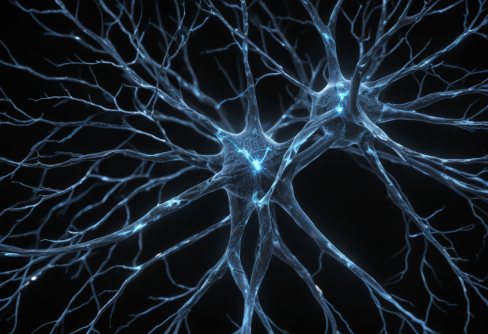
The reflex arc is the path along which excitation travels from the site of stimulation to the organ that produces the response. Its components are a sensory receptor, afferent neurons, interneurons (also called relay neurons) located in the CNS, efferent, or centrifugal, neurons, and effector organs such as muscles and glands.
Physiology in practice: the autonomic nervous system
The autonomic nervous system is divided into a sympathetic and a parasympathetic part. The sympathetic part prepares the body for physical exertion. It can speed up and strengthen heart contractions, widen the bronchi, affect blood vessels and direct resources to situations where the body has to act.
The parasympathetic part works largely in the opposite direction. It supports digestion, promotes contraction of the bladder muscles and relaxation of the sphincter muscles, and lowers heart rate. So what is actually going on? The body isn’t ready to sprint all the time. It also has to be able to recover, digest, sleep and build up energy.
The word trophic is also used to describe sympathetic influence. For example, a sympathetic nerve does not trigger heart contractions from scratch, because the heart works automatically. Stronger contractions can, however, result from a change in the functional state of the heart muscle, from increased excitability and contractility, and from metabolic processes running more efficiently.
The brainstem contains the reticular formation, which consists of nuclei scattered throughout the brainstem. Afferent nerve fibers arrive there from many regions. The reticular formation helps regulate sleep and wake cycles. Visual and auditory stimuli and mental activity keep us alert, while a monotonous signal can encourage drowsiness.
The importance of the brainstem shouldn’t be underestimated. The midbrain, pons and medulla oblongata form the link between the brain and the spinal cord. The nuclei of the cranial nerves are located there: of the 12, 10 leave the brain through the brainstem. The ventricles of the brain connect to the central canal of the spinal cord. Even minor damage to the brainstem can be life-threatening.
What physiology teaches us about movement
The spinal cord is not just a cable between the brain and the muscles. It also has its own reflex function. Through its nerve centers, spinal reflexes can occur independently, while the conduction function relays excitation to other nerve centers.
In the gray matter of the spinal cord, 2 anterior and 2 posterior horns are distinguished: the anterior horn, the posterior horn and the lateral horn. The anterior horns contain the cell bodies of the effector neurons, and the posterior horns contain the sensory nuclei. The white matter holds the ascending and descending pathways. Ascending pathways carry sensory impulses from the periphery to the spinal cord. Descending pathways carry efferent impulses from different parts of the brain to the motor cells of the spinal cord.
As nerve fibers leave or enter the spinal cord, they form the anterior (motor) and posterior (sensory) roots. In each intervertebral foramen, these join to form a spinal nerve. Before joining the anterior root, the posterior root forms a sensory ganglion, also called the spinal ganglion. Humans have 31 pairs of spinal nerves.
When you think about training, this explains a lot. Muscle work is not only about strength or willpower. A precise movement requires receiving sensory information, processing in the CNS, a motor response, balance, muscle tone and autonomic support. Honestly, every smooth squat or running stride is a small collaborative project involving the whole nervous system.
FAQ
What is the difference between the CNS and the peripheral nervous system?
The CNS consists of the brain and spinal cord and processes information. The peripheral nervous system connects the CNS with receptors, muscles, glands and internal organs.
Why does the body need reflexes?
Reflexes allow a quick response to a stimulus. Some of them are innate, while others develop through learning and repeated experience.
How does the autonomic nervous system affect training?
The sympathetic part helps you get ready for exertion, and the parasympathetic part supports recovery. You need both, because the body has to be able to work as well as calm down.
Author: EKFL
Source: WHO – physical activity.
Come and work out! ArtGym